Stress Echo Test: Purpose, Procedure & Results


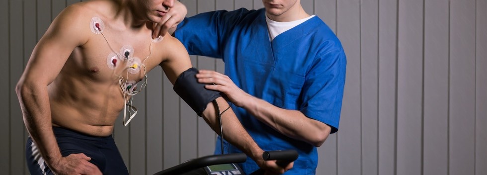
Table of Contents
In the dynamic world of cardiology, the Stress Echo Test has emerged as an essential tool. This fundamental procedure plays a vital role in assessing cardiac function and identifying a range of heart conditions. If you are seeking to deepen your knowledge and explore this topic, this blog article aims to provide a comprehensive understanding of the Stress Echo Test‘s purpose, procedure, and the invaluable information it offers.
This test is particularly beneficial in diagnosing coronary artery disease, heart valve disorders, and assessing overall cardiac function, enabling early detection and proactive management of cardiovascular issues. Throughout this article, we will unravel the step-by-step procedure involved in the Stress Echo Test, shedding light on the techniques employed to induce stress, the non-invasive monitoring methods, and the imaging techniques used to capture the heart’s response. Moreover, we will explore the significance of interpreting the test results, empowering both patients and medical practitioners with vital knowledge to make informed decisions regarding heart health management.
What is Stress Echocardiography?
Stress echocardiogram is a widely used non-invasive diagnostic test that plays a crucial role in the field of cardiology. This test provides valuable insights into the function and structure of the heart, helping healthcare professionals assess cardiac health and detect potential abnormalities.
The procedure involves combining two essential techniques: echocardiography and stress testing. Echocardiography utilises ultrasound to create detailed images of the heart’s chambers, valves, and blood flow, allowing physicians to visualise any structural irregularities or abnormalities. On the other hand, stress testing involves inducing the heart to work harder, either through physical exercise on a treadmill or by administering specific medications that simulate the effects of exercise.
During the stress echo test, the patient’s heart is monitored closely through electrocardiogram (ECG) and blood pressure measurements while the stress-inducing phase is underway. The imaging process captures the heart’s response under stress, providing valuable data on its ability to handle increased demands. This dynamic evaluation is particularly beneficial for identifying coronary artery disease, evaluating heart valve function, and assessing overall cardiac performance.
One of the significant advantages of stress echocardiography is its ability to diagnose heart conditions that might not be apparent during rest or standard echocardiography. This helps in early detection and on-time treatment, which can be critical for improving patient outcomes and preventing potential complications.
What does the echocardiogram stress test show?
Stress echocardiogram provides a comprehensive evaluation of the heart’s performance under stress conditions. During the test, real-time images of the heart are captured at different stages: before, during, and after the stress-inducing activity, which can be induced either through exercise or medications. These images offer valuable insights for cardiologists to assess various crucial aspects of the heart’s function.
One of the primary parameters evaluated during the stress echocardiogram is blood flow. By monitoring the blood flow in different areas of the heart, the test can identify potential blockages or reduced blood supply to certain regions, indicative of conditions like coronary artery disease. This critical information allows healthcare professionals to carry out accurate diagnoses, enabling prompt intervention and appropriate management.
Another vital aspect evaluated is heart wall movement. The stress echocardiography assesses how well the heart muscles contract and relax under stress, which can be indicative of various heart conditions, including hypertensive heart disease or heart valve disorders. Abnormalities in heart wall movement can provide essential clues for further investigation and targeted treatment plans.
Cardiac function is also closely examined during the stress echocardiogram. The test helps determine how effectively the heart pumps blood during stress, providing information on the heart’s overall health and performance. This assessment can aid in identifying heart function impairment, guiding physicians in formulating personalised treatment strategies.
The stress echocardiogram also serves as a vital tool for diagnosing a range of heart conditions, such as heart arrhythmias, heart failure, and other structural abnormalities. By capturing dynamic images of the heart’s response to stress, this non-invasive procedure allows for a comprehensive evaluation of cardiac health, enabling healthcare providers to deliver precise and tailored care to patients.
What happens during a stress echocardiography?
Below-mentioned are are the step-by-step procedure that you should expect during your stress echo test:
Procedure for Stress Echocardiography Test
- Patient Preparation: The stress echocardiography test begins with the patient’s preparation, where a medical technologist or sonographer explains the procedure in detail and addresses any questions or concerns the patient may have. The patient may be asked to refrain from caffeine and smoking before the test.
- Consent and Vital Signs: Before the test commences, the patient is asked to sign a consent form, acknowledging their understanding and agreement to undergo the procedure. The healthcare team records the patient’s vital signs, including blood pressure, heart rate, and electrocardiogram (ECG), to establish baseline measurements.
- Electrodes and Gown: The patient dons a comfortable laboratory gown while adhesive electrodes are affixed to the chest for continuous ECG monitoring. In some cases, a gentle abrasion technique is utilised to enhance electrode adherence.
- IV Line Insertion (if applicable): If the stress test requires pharmacological stress agents like dobutamine, an intravenous (IV) line is inserted into the patient’s arm to administer the medication.
- Ultrasound Gel Application: The medical technologist applies a gel to the ultrasound area, usually located on the left side of the chest. This gel ensures better ultrasound probe contact and improves image quality.
- Resting Echocardiogram: The medical technologist performs the resting echocardiogram using an ultrasound probe. This non-invasive imaging technique captures detailed images of the heart’s structure and function at rest.
- Stress Induction – Exercise or Pharmacological Stress: The patient is subjected to stress through either exercise or the administration of pharmacological stress agents like dobutamine. In exercise stress testing, the patient may walk on a treadmill or ride a stationary bike, with the intensity gradually increasing to reach the target heart rate. In pharmacological stress testing, medications are intravenously administered to simulate the effects of exercise on the heart.
- Dynamic Echocardiogram Imaging: While the heart is under stress, additional echocardiographic images are obtained at various intervals. These dynamic images capture the heart’s function and structure during increased cardiac demand.
- Post-Stress Imaging and Recovery: After reaching the target heart rate or completing the pharmacological stress, the patient rests, and another set of echocardiographic images is acquired. This phase allows the medical team to observe the heart’s recovery and assess its ability to return to a resting state efficiently.
- Expert Review and Reporting: A cardiologist reviews the captured images and provides a specialist opinion. A comprehensive report is then sent to the patient’s healthcare provider for further evaluation and treatment planning.
Who should have a stress echo test?
- Chest pain or angina: A stress echocardiogram is commonly used to assess patients with chest pain or discomfort to determine if it is related to reduced blood flow to the heart or other heart conditions.
- Suspected coronary artery disease (CAD): Patients with risk factors for CAD, such as a family history of heart disease, high blood pressure, high cholesterol, diabetes, or smoking history, may undergo a stress echo to detect and evaluate any potential blockages in the coronary arteries.
- Heart conditions: For individuals with known heart conditions, a stress echocardiogram can be useful to monitor the effectiveness of treatment, assess the progression of the disease, or identify any new heart problems.
- Assessment after a heart attack: After experiencing a heart attack, a stress echo can help evaluate the extent of damage to the heart muscle and determine the need for further intervention or rehabilitation.
- Preoperative evaluation: In some cases, individuals with existing heart conditions may require surgery. A stress echo can help assess their cardiac function and risk tolerance before undergoing the procedure.
- Fitness evaluation for specific activities: Some individuals, such as athletes or those with physically demanding jobs, may undergo a stress echo to assess their heart’s response to exercise and ensure they are fit enough for their specific activities.
Who should not have a stress echo test?
A stress echo test should not be performed on individuals with:
- Unstable angina or recent heart attack
- Severe aortic stenosis
- Uncontrolled arrhythmias
- Aortic dissection
- Severe heart failure
- Acute illness or infection
- Pregnancy
- Severe lung disease
NOTE: Always consult a healthcare professional for personalised guidance.
What do the test results mean?
Here are some possible interpretations of the stress echo test results:
- Normal: If the test is normal, then the heart is functioning well under stress, with good blood flow to the different regions of the heart. This suggests a low likelihood of significant coronary artery disease or other structural abnormalities.
- Abnormal: If the test is abnormal, it indicates various issues, such as reduced blood flow to specific parts of the heart, abnormal heart wall movements, or other abnormalities in cardiac function.
Some of the possible abnormal results can also be Coronary Artery Disease—where the coronary arteries are narrowed or blocked, restricting blood supply to the heart muscle, Valve Abnormalities—suggesting heart valve disorders, such as aortic stenosis, mitral valve regurgitation, or other valve-related issues or also Hypertensive Heart Disease—a condition caused by high blood pressure that affects the heart’s structure and function.
Based on the stress echo test results and other clinical factors, the healthcare provider will determine the appropriate course of action, which may include further diagnostic tests, lifestyle modifications, medication, or interventional procedures.
Conclusion
Stress echocardiography is a valuable non-invasive diagnostic tool in cardiology that can help in gaining critical information about cardiac function and detecting various heart conditions. The test’s dynamic nature allows for the assessment of heart performance under stress, enabling early detection and proactive management of cardiovascular issues.
For individuals seeking access to reliable and skilled healthcare providers, BP in Control offers a comprehensive online portal called “Find a Physician“. Through this platform, you can easily find the best physicians in your nearest periphery, including specialists in cardiology. Additionally, exploring BP in Control’s other blogs can provide further information on topics like hypertensive heart disease and related heart health topics, empowering individuals to take charge of their well-being and make informed decisions regarding their health. Remember, your heart health is in your hands, and BP in Control is here to support you every step of the way.
Disclaimer
The information contained in this article is to educate, spread awareness in relation to hypertension and other diseases to the public at large. The contents of this article are created and developed by BPinControl.in through its authors, which has necessary, authorisations, license, approvals, permits etc to allow usage of this articles on The Website. The views and opinions expressed in this article are views, opinions of the respective authors and are independently endorsed by doctors. Although great care has been taken in compiling and checking the information in this article, The Website shall not be responsible, or in any way liable for any errors, omissions or inaccuracies in this article whether arising from negligence or otherwise, or for any consequences arising therefrom. The content of this article is not a substitute for any medical advice. The Website shall not be held responsible or liable for any consequence arising out of reliance on the information provided in the article.
Comments (0)
No comments found.Add your comment